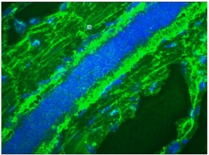
Mitotic Cells Mouse Monoclonal Antibody [Clone ID: 8B3G]
Specifications
| Product Data | |
| Clone Name | 8B3G |
| Applications | FC, IF, IHC |
| Recommended Dilution | Flow Cytometry: 1/50-1/100. Immunohistochemistry on Frozen Sections. Immunocytochemistry: 1/50-1/100 with avidin-biotinylated horseradish peroxidase complex (ABC) as detection reagent. Using immunocytochemistry, a combination of this antibody and BrdU (Cat.-No BM6048P) can distinguish and quantitate the four major fractions of the cell cycle. Not suitable for Immunoblotting. |
| Reactivities | Human, Zebrafish |
| Host | Mouse |
| Isotype | IgM |
| Clonality | Monoclonal |
| Immunogen | Total cell lysate of the Human bladder carcinoma cell line T24. |
| Specificity | This antibody detects Mitotic cells in Human samples. 8B3G strongly stains mitotic cells and can therefore be used in Flow Cytometric analyses of cell suspensions to detect the mitotic index. Together with a quantitative DNA staining procedure (e.g. propidium iodide) 8B3G clearly distinguishes these M-phase cells from cell at other stages of the cell cycle. Dynamic information can be obtained by combining BrdU incorporation with 8B3G staining, which can distinguish and quantitate the four major fractions of the cell cycle. |
| Formulation | PBS State: Purified State: Liquid purified IgG fraction Preservative: 0.09% Sodium Azide |
| Concentration | lot specific |
| Purification | Immunoaffinity Chromatography |
| Conjugation | Unconjugated |
| Storage | Store undiluted at 2-8°C for one month or (in aliquots) at -20°C for longer. Avoid repeated freeze-thaw cycles. |
| Stability | Shelf life: One year from despatch. |
| Background | The life cycle of a eukaryotic cell consists of various phases, two of which can morphologically and biochemically be identified. Firstly, during mitosis (M-phase), in which the cell divides into two identical daughter cells, chromosome condensation and spindle formation are microscopically visible. Secondly, in S-phase the DNA of a cell is replicated, a process that can be detected using biochemical techniques, such as the BrdU incorporation assay. In between the M- and S-phase two gap phases occur: the G1-phase, the gap between mitosis and the start of DNA replication, and G2-phase, the gap between completion of DNA replication and the onset of mitosis. From G1-phase a cell can leave the cell cycle and enter G0, a 'quiescent' phase. Regulation of the cell cycle predominantly occurs at three major control points, which govern the transition from G0 to G1, from G1 to S, and from G2 to M-phase. M phase itself is highly regulated, and is divided into five stages: prophase, prometaphase, metaphase, telophase and anaphase. |
| Reference Data | |
Documents
| Product Manuals |
| FAQs |
| SDS |
{0} Product Review(s)
0 Product Review(s)
Submit review
Be the first one to submit a review
Product Citations
*Delivery time may vary from web posted schedule. Occasional delays may occur due to unforeseen
complexities in the preparation of your product. International customers may expect an additional 1-2 weeks
in shipping.


 Germany
 Japan
Japan
 United Kingdom
 China
China


